题目:Compressed Sensing and Applications in X-ray Computed Tomography
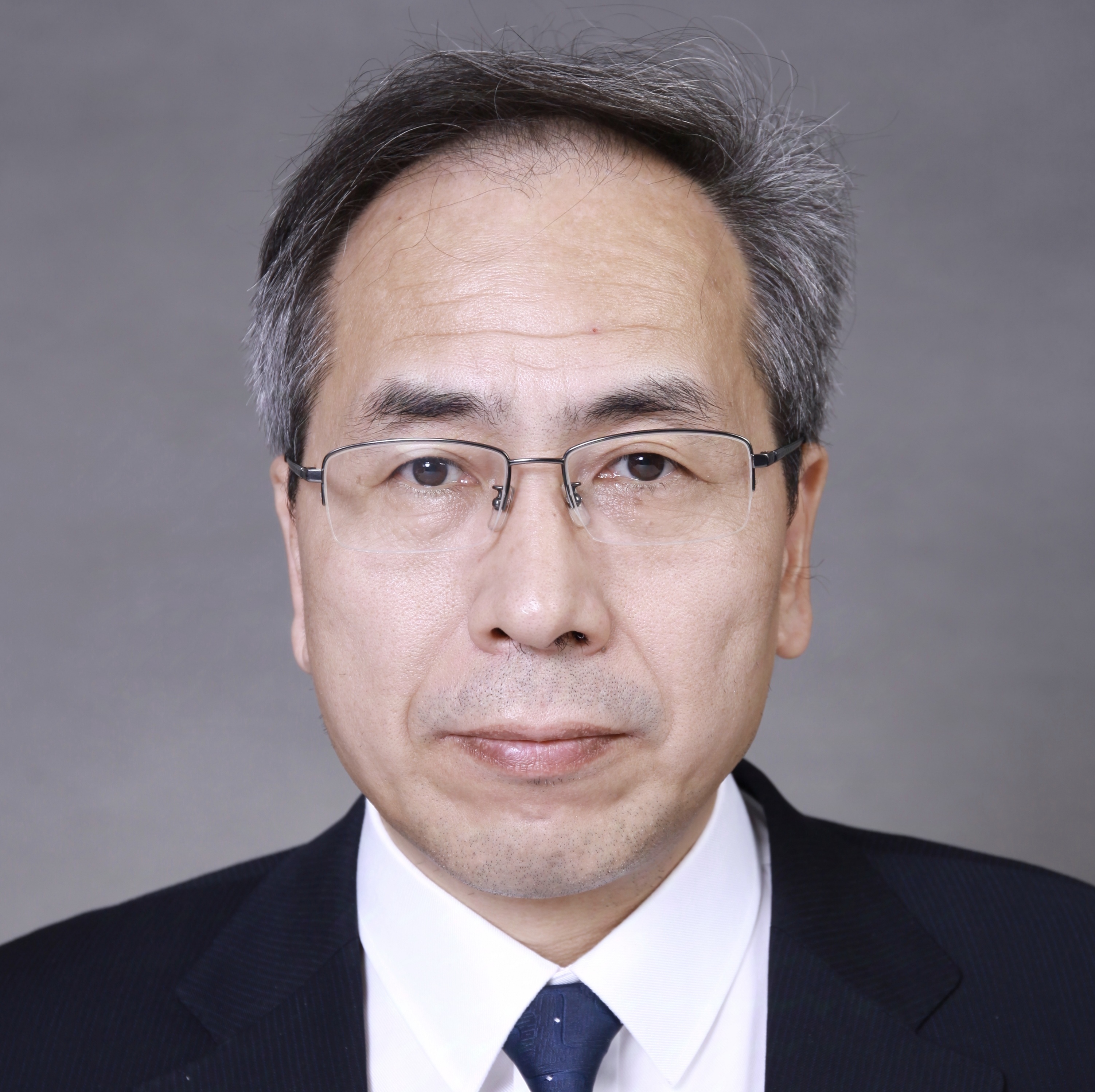
姓名:Guanghong Chen
单位:Department of Medical Physics and Radiology and the Director of X-ray
CT Imaging Center, University of Wisconsin-Madison
时间:6月19号(周二)上午10:00~11:30
地点:一号楼第五会议室(一楼多功能厅旁边)
摘要:
Compressed sensing (CS) has been an intriguing mathematical framework in information science. It extends the concept of image compression in image processing to physical acquisition process: i.e., If a digital image is compressible or sparse in some representation domains, we may have already acquired too much redundant information to generate the image. The CS theory intended to address the issue of how much data samples do we need to acquire to enable us to reconstruct the digital signal/image accurately provided that the image is sparse in some representation domain? It has been mathematically proven by the pioneers in CS theory that, under some conditions, one only needs to acquire vastly under-sampled data points for an accurate image/signal restoration. Namely, the number of data samples can be far fewer than that required by the conventional Shannon/Nyquist Sampling requirements. Since the birth of the CS theory, scientists have been worked individually or together to strive for practical applications of the CS theory in diversified scientific disciplines.
In this lecture, we will discuss how to incorporate the CS concept into the well-studied image reconstruction problem in x-ray computed tomography (CT). We will start with the formulations of the CT image reconstruction problem and outline the primary challenges in X-ray CT. Based on the challenges in CT, the original CS formulation was modified to incorporate a high signal-to-noise (SNR) prior image into the problem formulation. This image reconstruction framework has been referred to as Prior Image Constrained Compressed Sensing (PICCS). Given the practical constraint of reconstruction time must be limited to about a minute or so for an image volume with 512×512×512 voxels and also the constraint of radiation doses allowed for medical applications, innovations and advances from optimization community, computer science, and medical physics have been integrated to enable PICCS work for clinical applications. Finally, contributions form clinicians have been integrated to validate the PICCS reconstruction framework and contributions from medical device companies have also been pooled together to attempt for final products to improve people’s quality of life in future. In summary, using PICCS as an example, we intend to share our lessons and experiences in medical CT with the audience and hope to inspire more in-depth discussion and establish collaboration with other experts from multi-disciplines.
报告人简介:
Dr. Guang-Hong Chen received his BS from Beijing Normal University in 1991, one year earlier than his peer classmates. From 1991-1994, Guang-Hong worked at Cangzhou Teacher's College in Hebei Province. From 1994-1997, Guang-Hong conducted his graduate studies under the supervision of Prof. Mo-Lin Ge. From 1997-1999, he moved to United States to study semiconductor physics under the supervision of Prof. Michael Raikh and Prof. Yong-Shi Wu. Before he received his Ph.D from the University of Utah, Guang-Hong was elected as Graduate Fellow to conduct research under the supervision of Prof. Matthew Fisher. After that, he went back to Utah to spend two years with Prof. Yong-Shi Wu as a Research Associate. In the fall of 2002, Guang-Hong completely switched his research directions from theoretical physics to medical physics. He started as a Research Associate at the University of Wisconsin-Madison to initiate new research directions in x-ray CT. In early 2003, Guang-Hong was appointed as a tenured track Assistant Professor and the Director of X-ray CT Imaging Center. After 4 years, Guang-Hong received his early tenure and promoted to Tenured Associate Professor in 2007. In 2011, Guang-Hong was promoted to the Tenured Full Professor of Medical Physics and Radiology and the Director of X-ray CT Imaging Center. Currently, Guang-Hong is supervising more than 20 Ph.D students, Postdocs, Research faculty, Staff, and Clinicians to conduct research in almost all the x-ray related research directions in medical applications. His research was continuously supported by federal funding agencies and medical industries with more than $10M funding support in the past years and received more than $10M research equipment support from medical industries. He is on the editorial board of many internationally renowned journals such as the journal of Medical Physics, he also served as a permanent member of many federal and private foundations. Up to date, Guang-Hong published more than 120 scientific papers and had been granted more than 20 US and International patents.
所办公室
2012.6.18


